
|
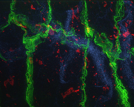
OMX-acquired image of a Drosophila blood-brain barrier (projection, early larval stage).
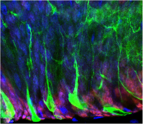
Green, Septate junctions. Red, gap junction (Innexin 1). Blue, Laminin. Confocal image showing a lateral view of a Drosophila ventral nerve chord (projection, early larval stage).
Green, neural stem cell membranes. Red, neurons. Blue, glia. Confocal image of a ventral view of a Drosophila ventral nerve chord (single slice, late larval stage).
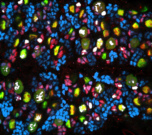
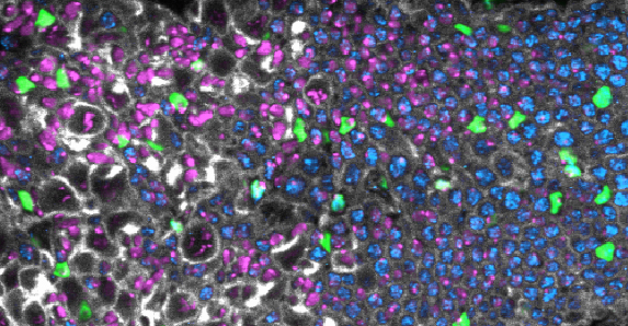
Green, neural stem cells. Red, fate determinant Prospero. Blue, neurons. White, phospho-histone 3. Tri-dimensional reconstruction of a confocal image showing Drosophila central brain and optic lobe (stack, late larval stage).
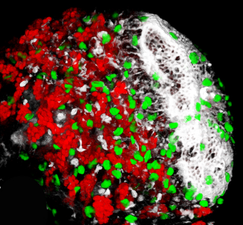
Green, glia. Red, neurons. White, actin. Confocal image of a ventral view of a Drosophila ventral nerve chord (single slice, late larval stage).
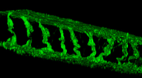
Green, glia. Blue, neurons. Magenta, nuclei (DAPI). White, actin. Tri-dimensional reconstruction of a confocal image showing a Drosophila blood-brain barrier (stack, lateral view, early larval stage).
Green, blood-brain barrier membrane. |
